|
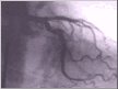
The current gold standard for identifying coronary artery disease and validating aortal and mitral valve disease is the coronary angiogram. The angiogram is often performed in conjunction with a cardiac catheterization. While these terms are frequently used synonymously, they actually refer to different tools. In a coronary angiogram, the physician introduces a small tube into the arterial system, typically through an artery of the groin or the arm. Using X-ray guidance, the physician guides that tube to the origin of the arteries that feed the heart. A dye is then injected through the tube to enter and flow through the coronary arteries. If needed, the dye may be injected directly into the left ventricle, aorta, or pulmonary arteries. The distribution of that dye enables the physician to directly visualize the coronary arteries, which remain invisible using a standard X-ray approach.
The angiogram reveals the presence or absence of coronary arteries, whether the arteries remain open or closed, and any indications of early arterial disease that may eventually significantly limit blood flow. Some blockages have specific properties that put so-called vulnerable plaques at risk. Vulnerable plaques may abruptly rupture, closing an artery that had not experienced any previous interruption of flow. A significant minority of patients, however, appear to progress gradually from slightly narrowed arteries to severe blockages that interrupt the arterial flow. These patients typically experience symptoms of angina or heart attacks from their blockages.
With the development of noninvasive diagnostic tests such as MRAs or CT angiograms, physicians will probably increasingly use angiography only for therapeutic applications such as balloon angioplasty, rather than as a diagnostic technique. In a balloon angioplasty, the physician winds the catheter to the blocked portion of the coronary artery. Once there, the physician inflates a balloon located at the tip of the catheter, pushing open the vessel. In addition, a stent or mesh framework may be placed in the blockage, permanently propping open the artery. Interventional cardiology may replace surgery for some patients as it can now be used to close congenital defects (PFO/ASD) and repair or replace aortic or mitral valves. While physicians may turn to noninvasive alternatives to initially diagnose arterial disease, the cardiac catheter will likely remain a significant treatment modality for some time to come.
 
|