|
The term echocardiography stems from the "echoes" of the sound waves that are captured to create the "cardiograph," or picture of the heart. Echocardiography relies upon nonradioactive and noninvasive ultrasound technology to image the heart. Sound waves are transmitted to the heart from a device called a transducer, which is applied to the skin. Once the waves reach the heart, they bounce back across the skin and are recorded by the transducer. Echocardiography currently offers the only truly comprehensive tool that can accurately diagnose all types of heart disease in a single examination, including congenital defects.
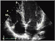
The physician places the transducer on the chest wall and directs the sound waves between the ribs to record different locations within the heart. The resulting images can be captured at various speeds and displayed on a screen for immediate viewing during the examination. The advent of digital technology enables us not only to acquire increasingly clear images, but also to store these images electronically—recalling them for quick side-by-side comparisons on a computer screen with the click of a mouse.
Intravenous contrast injection of sterile saline can be used to detect congenital defects that may lead to stroke. Images can be enhanced with nonradioactive intravenous contrast agents but this is not usually necessary.
A resting echocardiogram identifies both the anatomy and function of the heart. Regional differences in muscle function may appear in the echocardiograms of patients who have previously experienced heart attacks or who have advanced heart disease. When combined with exercise, echocardiography can help physicians to compare the response of the heart muscles to stress, both in terms of overall function and in terms of regional differences in capability. This comparison can offer physicians very important insights into which patients face increased risk for heart attack or heart failure—helping to identify those patients who require more aggressive interventions such as angioplasty or surgery.
The Doppler technique, another adaptation of ultrasound testing, depicts blood flow. It can accurately diagnose the location and degree of valvular blockages and leaks, as well as the presence of heart muscle (pericardial) disease. Physicians can also use stress echocardiograms to monitor the progress of severe valvular disease or heart muscle disease by examining indicators of cardiac performance. Finally, digital echocardiography can help physicians determine the optimal timing and technique for a surgical intervention, if necessary.
 
|