|
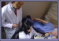
Physicians can employ a variety of techniques to diagnose heart disease. As a general introduction, we will first look at some historical approaches, and then turn to specific, technology-based diagnostic techniques. Every tool we use has both a limit and a value, which we think of in terms of sensitivity and specificity. Initially—and very importantly—we find ourselves still anchored in the historical tradition of conducting a careful interview, called a history, and a physical examination. Often the physician can diagnose the disease and determine a proper course of management with a fair degree of certainty using these two tools. Physicians acquire the necessary skills through repetition and training, in medical school and beyond.
To offer our patients the best possible care, we also have to apply the right diagnostic tool to the specific case. I will outline a number of the diagnostic tools available today, including electrocardiograms (EKGs), angiograms, nuclear scans, computerized-axial tomography (CAT or CT scans), magnetic resonance imaging scans (MRIs), magnetic resonance angiogram scans (MRAs), and digital echocardiograms. Physicians typically rely on the history, physical, and perhaps an electrocardiogram to make the initial probable diagnosis. Based on that initial impression—which takes into account the patient's risk factors for coronary artery disease, heart attack, heart failure, and stroke—we then pick the right tool to confirm our findings and determine the best possible treatment plan.

|