|
Pathology
In aortic stenosis the tricuspid, or three-part, aortic valve becomes stenosed, or narrowed. Typically, this narrowing stems from the development of calcifications. Among older patients, particularly for 70- to 90-year-olds, this constriction can represent a natural part of the aging process. In the younger population—most commonly for 40- to 50-year-olds, but sometimes even for teenagers—this narrowing more often results from a congenitally abnormal valve. Approximately one percent of infants are born with a bicuspid, or two-part, aortic valve instead of the normal tricuspid construction. Because of the flow characteristics across their edges, bicuspid aortic valves develop earlier scarring and calcification. Rheumatic heart disease is also a risk factor for the development of aortic and mitral stenosis, although rheumatic fever is now rare in the U.S. due to early diagnosis of strep infections and effective antibiotic therapy.
Once the valve becomes so narrowed that the opening is inadequate for normal forward blood flow, the heart will increase its muscle mass, in a condition known as hypertrophy. By building muscle, the heart will create higher pressures in the left ventricle, seeking to force the valve to open wider and deliver a greater blood flow. Eventually, the heart will reach a pressure differential of 50 to 100 millimeters of mercury between the left ventricle and aorta—the limit of its ability to compensate for the faulty valve. Patients who have this condition may experience shortness of breath, chest pain, heart failure, fainting, or even sudden death.
Diagnosis
The symptoms of aortic stenosis usually include chest pain, particularly with effort or exercise, shortness of breath, fainting, or heart failure. Physicians often detect aortic stenosis by picking up the presence of a murmur through a stethoscope. The murmur from a stenotic aortic valve has specific characteristics; to a limited degree, physicians can even estimate the severity of the stenosis through the physical examination. (Remember, a murmur means a doctor hears blood flow with a stethoscope, which is not always a sign of disease.)

| Physicians can detect aortic stenosis by picking up the sound of a murmur. |
|
|
Get the Flash Player to see this player.
|
Currently, echocardiography (ultrasound) is the definitive technique for diagnosing aortic stenosis. This tool allows the physician to visualize the valve and its movement, measuring the size of the opening as well as the amount of pressure built up behind the valve. The approach has been validated against the previous, more invasive standard of angiography for the management of aortic stenosis.
The EKG remains a tool in estimating the severity of the narrowing, as well as in establishing the proper timing for a possible surgical replacement. Severe aortic stenosis can be indicated by the development of an increasing amount of voltage on the patient's EKG. This increase often reflects hypertrophy, the thickening of the heart muscle to compensate for the narrowed valve.
Treatment
Valve replacement surgery has traditionally been the treatment technique of choice for aortic stenosis patients. Efforts to use balloons to dilate the valves, known as valvuloplasty, have improved symptoms and performance only for a very short time period. Since valvuloplasty has a high rate of recurrence within six to 12 months, most patients elect to undergo valve surgery.
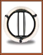
Aortic stenosis surgery can be performed safely and quickly, offering patients a durable result. In aortic valve surgery, the surgeon opens the aorta, removes the defective aortic valve, and replaces the patient's valve with either a tissue valve (usually a pig valve) or a mechanical valve. Most patients elect to receive a mechanical valve, which is manufactured to approximate the normal operation of the patient's own heart valve. The average tissue valve lasts only for seven to 10 years. However, I have had patients whose original valve implant is still working after 16 to 17 years.
While mechanical valves are more durable, they are also more likely to create blood clots on the valve's surface. These clots may break free and cause complications, such as strokes. To avoid clots, physicians require mechanical valve patients to take a powerful blood thinner, usually coumadin. Blood thinners decrease the risk of clotting, but increase the risk of bleeding. The use of coumadin and other blood thinners, therefore, is a concern for patients who have bleeding tendencies from other conditions, or who are unreliable in their use of medications. Theoretically, mechanical valves can last a lifetime. They have been known to last for more than twenty years.
A new experimental option is to use a catheter to implant a stent valve over the existing stenotic valve in combination with valvuloplasty. Stent valve bioprosthetic replacement could take the place of conventional open-heart surgery techniques.  
|