| |
 |
 |  |
|
High homocysteine levels can serve as a marker for coronary artery disease.
   | |
|
|
|
|
As part of preventing heart disease, we need to screen more people for the presence of arterial plaque. The first order of business is to look at serum cholesterol, the level of cholesterol in the bloodstream. Physicians can associate that total cholesterol count with an increased or decreased risk of heart disease. However, we can now break that overall finding down into more specific counts of high-density lipoprotein (HDL), the so-called good cholesterol, and low-density lipoprotein (LDL), or bad cholesterol. From a given level of HDL or LDL, physicians may be able to identify a definable risk in patients. HDL and LDL subparticle analysis may more clearly define patients at risk as well as pinpoint potential supplementary drug therapies for those whose cholesterol levels are not completely controlled by statins alone.
As we continue to examine cholesterol subfractions, we are becoming increasingly sophisticated about which particles are the most active for disease progression and disease prevention. For example, physicians now test for triglycerides, which the body manufactures from carbohydrates and fats. High triglyceride levels are associated with an increase in cardiac risk. We are also learning how to better influence triglyceride levels, using diet, exercise, vitamins, and prescription medicines.
Physicians can turn to techniques that identify plaque noninvasively—or, as one of my patients said, we can try to find the plaque before it finds you. We conduct a careful history and physical exam, looking for clues about the possible presence of disease. We listen for arterial noises and check for asymmetric blood pressure readings between the arms and legs. We examine an electrocardiogram for evidence of previous silent heart attacks or of irregular heart rhythms, both of which we associate with artery disease.
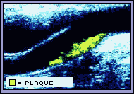
We increasingly depend on noninvasive imaging techniques to look inside the body to establish the presence or absence of plaque. Carotid (neck) artery ultrasound allows us to envision the inside of the arterial wall at a very detailed level, measuring differences of less than one millimeter of thickness. Using this technology, we can look for any thickening of the cells lining the arterial wall, an early sign of plaque, as well as the presence of calcifications and the degrees of narrowing. The presence of plaque in the carotid arteries indicates its presence elsewhere in the body, particularly in the coronary arteries. It also indicates an increased risk for stroke and heart attack.
Physicians can directly treat blockages in the carotid, coronary, and peripheral arteries using balloon angioplasty, stents, and surgical techniques. By following the increase or reduction of plaque in these arteries, physicians can track the general progress of their patients, noting improvements in response to lifestyle changes and medical therapies. As an alternative approach, CT scanning can directly detect coronary artery calcium, a marker for cholesterol-rich blockages particularly among men, who typically develop calcifications earlier than women.
 
|